

版权说明:本文档由用户提供并上传,收益归属内容提供方,若内容存在侵权,请进行举报或认领
文档简介
Chapter
8The
disease
of
cardiovascularsystem8.1
atherosclerosis
(
AS
)z
characterized
by
intimal
lesions
calledatheroma
or
fibrofatty
plaques
that
protrudeinto
the
lumen,weaken
the
underlyingmedia,and
undergo
a
serious
of
complicationsz
Primarily
affects
elastic
arteries
(aorta,carotid,
iliac-arteries)
and
large
and
mediusized
muscular
arteries
(coronary
andpopliteal
arteries)8.1.1
epidemiology
and
risk
factorshyperlipemia
/
hypercholesterolemia:①
LDL/LDL-C
,VLDL,
triglycerides,cholesterol→positive②
HDL/HDL-C,
apoA-I
→negativehypertension:→early
and
severesmoking:→endothelial
damage、CO↑→
PDGF↑,SMC
proliferation
emigration→
ox-LDLDiabetes
and
hyperinsulimemia:hyperglycemia→LDL
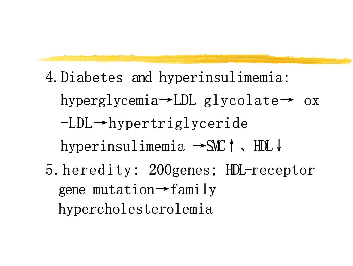
glycolate→ox-LDL→hypertriglyceridehyperinsulimemia→SMC↑、HDL↓heredity:
200genes;
HDL-receptorgene
mutation→familyhypercholesterolemia6.
Age
and
sex:age:
age↑→ASfactors
changesarterial
wall
proliferativechangesex:
estrogen→cholesterol↑postmenopausal
female=maleOthers:Obesity
,
stressful
life
type
,
hypoxia
,
dielackofVit.
C
,
infection
ofbacteria
,
viruLack of
exercise
:Intake
of
alcohol
:Protective
role
for
moderate
intake
ofalcohol
:
HDLA
large
amount
of
intake
of
alcohol:hypertension,
cerebral
hemorrhage8.1.3
pathologic
changeselastic
arteries:
aorta,
carotid,
iliaclarge
and
medium-sized
muscular
arteriecoronary,popliteal
A1.
favorite
site:lower
abdominal
aorta,
coronary
A,popliteal
A,
the
descending
thoracic
athe
internal
carotid
A,
the
circle
of
wWhat
does
the
atherosclerotic
vessel
look
lThe
lesion
of
atherosclerosis
is
not
one
spentitybut
a
spectrum
of
arterial
changesincluding
:□Atheromatous
fibro-lipid
plaques
=
fibroplaque
=
fibrotheroma□Fatty
streaks
=
intimal
xanthoma□Intimal
cushion
lesions
=
intimal
mass
lebasic
pathologic
changes1.
fatty
streak:
the
early
lesiongross:tiny
round
or
oval
flat
yellow
dotsarranged
in
rows,
coalesce
to
form
astreak,
1-2mm,
slightly
raised,particularto
theaortic
valve
regionLM:
a
collection
of
foam
cellsfatty
streak:
multipleyellowflatspots(fatty
dotFatty
streak
(sudan
red)(3)
formation:lipid↑→engulfed
by
Mφ,
SMC
→foam
cell(two
sources)(4)
result:reversiblecould
be
seen
in
infants
and
children2.
fibrous
plaque:gross:irregular
grey-white
raisedplaqueLM:
fibrouscap,
foam
cell,lipids图8-5
主动脉粥样硬化主动脉后壁见黄白色纤维斑块lumenFibrous
capLipid
core,clefMassontrichrome(胶原纤维-蓝;肌、纤维素-红)Media
thinCalcification,neovascular3.
Atheromatous
plaqueor
atheroma(hallmarkof
AS)z
gross:white/whitish
yellow/
yellowirregularelevated
plaque(2)
LM:①
surface:
fibrous
cap(hyaline
degenerationofcollagen,
SMC
embed
in
extra
cellularmatrix)②
necrotic
center:
amorphous
necrotic
materia(cell
debris,lipid,cholesterol
crystals,
casurrounding:
granulation
tissue,
lymphocyte+foam
cells③
media:
atrophy→thinComplicated
lesions:on
the
basis
of
fibrous
plaque
and
atheromatouhemorrhage
within
plaque:acute
obstruction
of
A(
coronary
A
→myocardial
infarction)focal
rupture:lower
abdominal
aorta、iliacarteries、tempArupture→ulceration→thrombosis↓atheromatous→embolism→infarct图8-8腹主动脉粥样硬化斑块内出血粥样斑块纤维帽和坏死物质之间可见大量红细胞,坏死物内可见针形“胆固醇结晶裂隙”图8-9腹主动脉瘤肾动脉下腹主动脉粥样硬化伴局限性明显扩张,动脉瘤形成。thrombosis:obstruction→infarctioncalcification:harden,
elasticityaneurysm
formation:rupture→hemorrhageAS
focal
disruption,
thrombomosiPslaque
ruptureThe
atheromatous
plaque
and
the
dynamics
of
plaquestability(三)Lesions
in
organs
&
clinical
features1.
aortic
AS:site:abdominal
aorta>thoratic
A>aorta arch>ascending
Alesions:atheromatous
and
secondarychangesulceration、calcification、hemorrhage,abdominal
aortic
aneurysm→Rupture→Death2.
Coronary
AS:coronary
heart
disease3.Carotid
A、Cerebral
A
ASsite:basilar
A,middle
cerebral
A,Williscirclelesion①ischemicatrophy②infarction:thrombosis→occuludetemporal
lobe,
caudate
nuclears,lenticularnucleus,thalamus③
hemorrhage:AS→aneurysm
rupture图8-10
大脑基底动脉粥样硬化
图8-11脑软化↑示动脉粥样硬化斑块
↑示筛状软化灶Renal
A
AS:sites:ostia
ofrenal
A
majorbranch,proximal
segmentlesion
:
infarctor
hypertension
AS
of
extremities:narrowing→claudication→thrombosis→gangreneSmall
intestine
AS:
infarction8.2
pathogenesis1.
Response
to
injury
theory
andinflammation
theory:---
Current
view
of
the
pathogenesis
of
ASAS
plaque
as
the
response
of
endothelial
celto
damaging
factorsdamage
factors:mechanical
denudation、
LDL、hypercholesterolemia、immunecomplex、ox-LDL、smokingz
Central
to
this
thesis
are
the
followingeventsRoles
ofLipidsEndothelial
injurySmooth
muscle
proliferationMacrophage(Mφ)zz
①
Role
of
lipidsIncrease
endothelial
permeabilityImpairendothelial
functionOX-LDLingested
by
Mφ→foaming
from
cellschemotactic
for
circulating
monocytesincrease
monocyte
adhesionstimulate
release
of
growth
factorschemotactic
to
endothelial
cells
(EC)smooth
muscle
cells
(SMC)②
Role
of
EC
injury:
initial
lesionMechanical
injurySmokingRisk
Factors
HypoxiaHyperlipidemiaHypertensionEC
injuryNon-denuding
EC
dysfunctionEC
denudingIncreased
EC
permeabilityEnhanced
monocyte,
platelet
adhesionRelease
GF→SMC
proliferationz
③
role
of
macrophage:
key
stepy
Platelet
,
EC,
Monocyte,
SMCGrowth
Factors
(PDGF,FGF,etc)emigrationproliferationadhere→emigrant
subendothelially→
foam
cellsMatrixGF→SMC↑z
④
role
of
SMC
proliferation:GF
→
SMC
proliferation
in
media→
emigration→intimaaccounting
for
the
progressive
growth
of
ASphenotype
change(constrictive→synthesis)surface
LDL
receptor→foam
cell
(SMC
derived)collagen
,
elasticfiber
protein,
proteoglycansinteraction→formation
of
AS
plaque2.
Lipid
infiltration
theory:hyperlipemia↓EC
damage→permeability↑→lipoprotein
deposit→Mφcleanup↓SMC↑↓atheroma3.
Smooth
muscle
mutagenic
theory①SMC
in
AS
plaque→monoclone②
etiology:chemical
mutagenic
agentsvirus4.
Macrophage
receptor
defect
theory(1)normal①cell
membrane:
LDL
receptor→combine
withLDL→engulf→intracellular②
intake→LDL
receptor→change
with
the
needof
cholesterol(2)
AS:
LDL
receptors↓↓→LDL
cleaning↓→LDL
in
blood↑(2)
ox-LDL①
not
be
recognized
by
nativeLDL
receptor②
engulf
mediate
by
scavenger
recept(Mφsurface)→foam
cell③
chemotactic
to
monocytes↑④chemotactic
to
EC、SMC↑GF↑8.2
coronary
AS
and
coronaryheart
diseasecoronary
atherosclerosisdistribution:①left>right,large
branch>small
branch,proximal
segment>distant
segment②
left
anterior
descending
coronary
A>rightCA>left
CA>left
circumflex
CAThe
progression
of
myocardial
necrosis
after
coronary
a.RCALCXLADfeatures:cutfacecrescent
plaquemyocardial
side,eccentric
stenosin4
grades:Ⅰ
<
25%
;
Ⅱ
26-50%;
Ⅲ
51-75%;
Ⅳ
>76%ischemic
heart
disease
(IHD)8.2.2
coronary
heart
disease
(CHD)1.
CHD:
ischemic
heart
disease(IHD)–
agroup
of
closely
related
syndromeresulting
from
myocardial
ischemia2.etiology
and
pathogenesis:①insufficient
blood
supply:
AS、sp②
increase
in
cardiac
energy
demand:BP↑↑,overworked,excite→work
load
of
heart↑An
imbalancebetween
the
perfusion
anddemand
of
the
heart
for
oxygenated
blooangina
pectorisz
1.
angina
pectorisA
symptom
complex
of
IHD
characterized
byparoxymal
and
usually
attacks
of
substernal
oprecordial
chest
discomfort
(constricting,squeezing,
chocking
or
kinfelike)cardiac
oxygendemand↑↑acute、temporarily,comparatively
coronary
Ablood
supply2.
clinical
typesstable
or
typical
angina:reliveby
rest
or
nitroglycerin,
chronicstenosing
coronary
AS(>75%)prinzmetal
or
variant
angina:episodic
angina,
occure
at
rest,
duecoronary
spasm,
ECG:S-T
elevated,unrelated
to
BP,
rate,
activity,vasodilators
respond
promptly(3)unstable
or
crescendo
angina:occure
with
progressively
increasingfrequencytend
to
be
of
more
prolonged
durationInduced
by
disruption
of
an
AS
plaque
witsuperimposed
partial
thrombosis
andembolization
or
vasospasm
(preinfarctiangina)myocardial
infarction,(MI)concept:acute
ischemic
necrosisdue
to
severe
sustainedischemia,heart
attacktypes:95%
LV,
transmural
orsubendocardial
infarction(1)subendocardial
myocardial
infarctio①
concept:an
area
of
ischemic
necrosis
l
to
the
inner
1/3,
at
most
½
of
the
ventric
wall
(papillary
muscle,
trabeculae
carn②
lesion:multiple
different
in
size
focnecrosis③
circumferential
infarction:entire
LV
endocardium(2)transmural
myocardial
infarctiontypical
infarction:①
the
ischemic
necrosis
involves
the
fulnearly
full
thickness
of
the
ventriculawall
in
the
distribution
of
a
singlecoronary
artery
>2/3→thick
layer
infarz②
favorite
sites:LAD
40-50%Anterior
wall
of
LV
nearapex,anterior
portion
ofventricular
septrm,
apexcircumferentiallyRCA
30-40%Inferior-posterior
wall
of
LV,posteriole
portion
ofventricular
septum,
inferior-posterior
RV
free
wallLCX
15-20%Lateral
wall
of
LV
exceptat
apex3.lesion:anemic
infarct,yellow
or
gray-red,dryirregular12hr
naked
eye½-1hr
EMz4.Lab:z1/2hr:
glycogen↓z6-12hr:myoglobin
↑z24hr:
GOT,LDH,GPT,CPK↑gross½-4hr4-12hrOccasionally
dark
motting12-24hrDark
motting1-3dMotting
with
yellow-tan
infarct
center3-5dHyperemic
border,
center
yellow-tan
softing7-10dMaximally
yellow-tan
and
soft
with
depressed
red-tan
margins10-14dRed-gray
depressed
infarct
borders3wGray-white
scar,
progressive
from
border
to
coreof
infarct>2mScarring
completeLM½-4hrVariable
waviness
of
fibers
at
border4-12hrBeginning
coagulationnecrosis,edema,hemorrhage12-24hrOngoing
coagulation
necrosis,
myocytehypereosinophilia,
marginal
contraction
bandnecrosis1-3dCoagulation
necrosis,with
loss
of
nuclei
andstriation,
interstitial
infiltrate
of
neutrophils3-7dBeginning
disintegratin
of
dead
myofibers,
earlyphagocytosis7-10dWell-developed
phagocytosis
of
dead
cells,earlyformation
of
fibrovascular
granulation
at
margin10-14dWell-estabished
granulation
and
collagendeposition2-8wIncreased
collagen
deposition>2mDense
collagenous
scar图7-14心肌梗死心肌梗死48h后,心肌细胞核消失,肌浆变成均质细颗粒状,横纹几乎消失。扩大的心肌间隙中可见多量嗜中性粒细胞浸润。图7-15心肌凝固性坏死(肌浆凝集)梗死的心肌纤维肌浆内可见明显增厚的波浪状横带(收缩带)图7-16
心肌梗死心肌梗死7天后,心肌细胞核几乎消失,肌浆变成红染无结构。可见增生的肉芽组织。图7-17急性心肌梗死左心室前壁线形破裂5.complication①
myocardial
rupture:acute,severe,1-3days
or
1week(within
2weefavorite
siteresultVentricular
free
wallHemopericardium
andcardiac
tamponadeventricular
septumLeft-to-right
shuntpapillary
muscleSevere
mitral
regurgitation②
ventricularaneurysm:a
late
complication,from
a
largetransmuralanteroseptal
infarct,paradoxicallybulgesduringsystole③mural
thrombos
温馨提示
- 1. 本站所有资源如无特殊说明,都需要本地电脑安装OFFICE2007和PDF阅读器。图纸软件为CAD,CAXA,PROE,UG,SolidWorks等.压缩文件请下载最新的WinRAR软件解压。
- 2. 本站的文档不包含任何第三方提供的附件图纸等,如果需要附件,请联系上传者。文件的所有权益归上传用户所有。
- 3. 本站RAR压缩包中若带图纸,网页内容里面会有图纸预览,若没有图纸预览就没有图纸。
- 4. 未经权益所有人同意不得将文件中的内容挪作商业或盈利用途。
- 5. 人人文库网仅提供信息存储空间,仅对用户上传内容的表现方式做保护处理,对用户上传分享的文档内容本身不做任何修改或编辑,并不能对任何下载内容负责。
- 6. 下载文件中如有侵权或不适当内容,请与我们联系,我们立即纠正。
- 7. 本站不保证下载资源的准确性、安全性和完整性, 同时也不承担用户因使用这些下载资源对自己和他人造成任何形式的伤害或损失。
最新文档
- 山东省济宁鱼台县联考2024-2025学年初三下学期中考模拟考试语文试题(文史类)试卷含解析
- 山东省惠民县联考2025年初三中考仿真模拟卷(一)化学试题含解析
- 湖南省益阳市桃江第一中学2024-2025学年高中毕业班历史试题学科备考关键问题指导系列历史试题适应性练习(一)含解析
- 四川省成都市实验中学2024-2025学年高三第二次模拟考试试卷物理试题含解析
- 扬州大学《食品试验设计与统计分析实验》2023-2024学年第一学期期末试卷
- 徐州幼儿师范高等专科学校《医疗健康商务沟通》2023-2024学年第二学期期末试卷
- 内蒙古化工职业学院《视频内容传达》2023-2024学年第二学期期末试卷
- 湖北省学业考:专题二匀变速直线运动的研究复习试卷2025届高考原创押题卷(2)生物试题试卷含解析
- 内蒙古工业职业学院《统计计算与软件实验》2023-2024学年第一学期期末试卷
- 浙江省绍兴市诸暨市2025届数学四下期末统考模拟试题含解析
- 军事科技现状及未来发展趋势分析
- 人教版数学五年级下册分数比较大小练习100题及答案
- DB21-T 3031-2018北方寒区闸坝混凝土病害诊断、修补与防护技术规程
- JJF(新) 116-2023 微机盐含量测定仪校准规范
- 创伤性硬膜下出血的健康教育
- 光电编码器课件
- 马原演讲之谁是历史的创造者
- 《人类征服的故事》读后感
- 硫酸艾沙康唑胶囊-药品临床应用解读
- 学生社交技巧与人际关系的培养
- DLT817-2014 立式水轮发电机检修技术规程

评论
0/150
提交评论